Direct Anterior Approach & Patient Specific Technology
Total Hip Replacements
Years of evolution and refinement
Combines the accuracy of 3D planning with respect for surrounding soft tissue
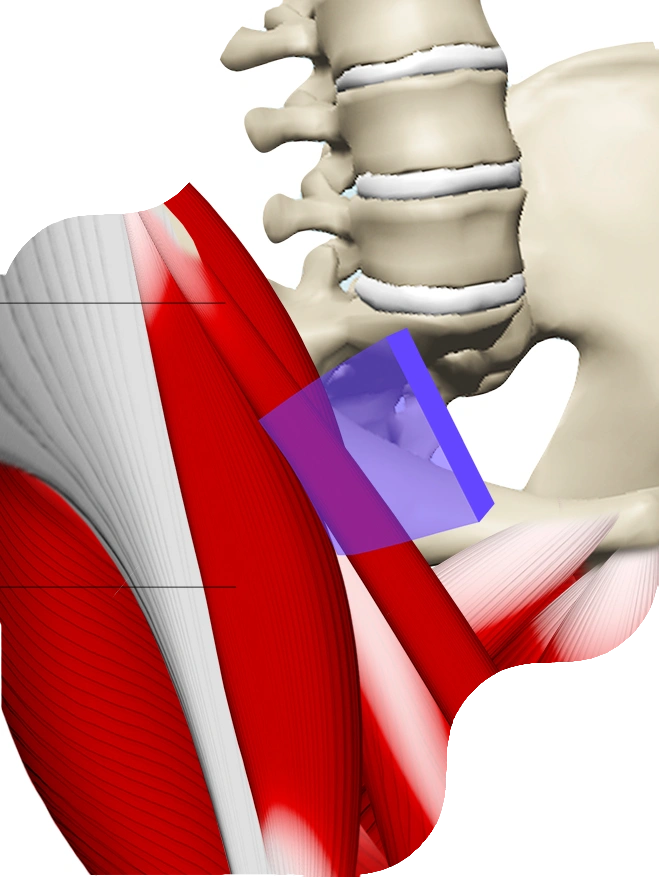
The direct anterior approach is a muscle sparing procedure

3D Imaging
Prior to surgery, a 3D scan is performed so that the surgery can be performed virtually. We now know the size, shape and position for each persons individual anatomy weeks before we enter the operating theatre.

Patient Specific
A custom made “jig” is created for each and every patient, which is a perfect fit with their anatomy – enabling accuracy during surgery for preparation of the bone.

Intra-operative checks
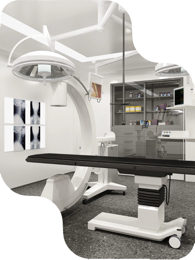
Very few methods allow intra-operative imaging during a hip replacement. A special table allows intra-operative Xray scanning that confirm accuracy.
DIRECT ANTERIOR APPROACH SURGERY
Respecting soft tissue structures

How its Performed
A truely internervous and intermuscular hip replacement procedure performed.
A 5-6cm incision is placed on the front of the hip (the hip is closest to the front of the body), and the muscle layers are moved aside rather than being detached. This affords significant stability to the joint after, as there are no tendons or muscles that need to reattach back to bone.
REVISION RATE
%
The Australian registry shows that Total Hip Replacements have an excellent survival. Approximately 8% of all hip replacements have been revised by the 20 year mark.
SUCCESS RATE
Hip replacements are one of the most successful operations with appriximately 95% of patients saying it was good or excellent.
How is the direct anterior approach different
The direct anterior approach is not a “new” approach – it has been used for decades. It has been a rapidly growing method for hip replacement surgeries over the last 20 years.
Accuracy
Using patient specific technology, measurements are done in 3 dimensions and custom made jigs are created to ensure accurate bony preparation
Soft tissue sparing
The surgery is performed without cutting or detaching muscles, tendons or ligaments, unlike most operations.
Imaging during surgery
The ability to perform an Xray during surgery is invaluable, as it allows an assessment prior to the final implant is placed. This further enhances accuracy.
Heavily researched
The direct anterior approach has been heavily researched over the years, and has been our preferred method for over 10 years.


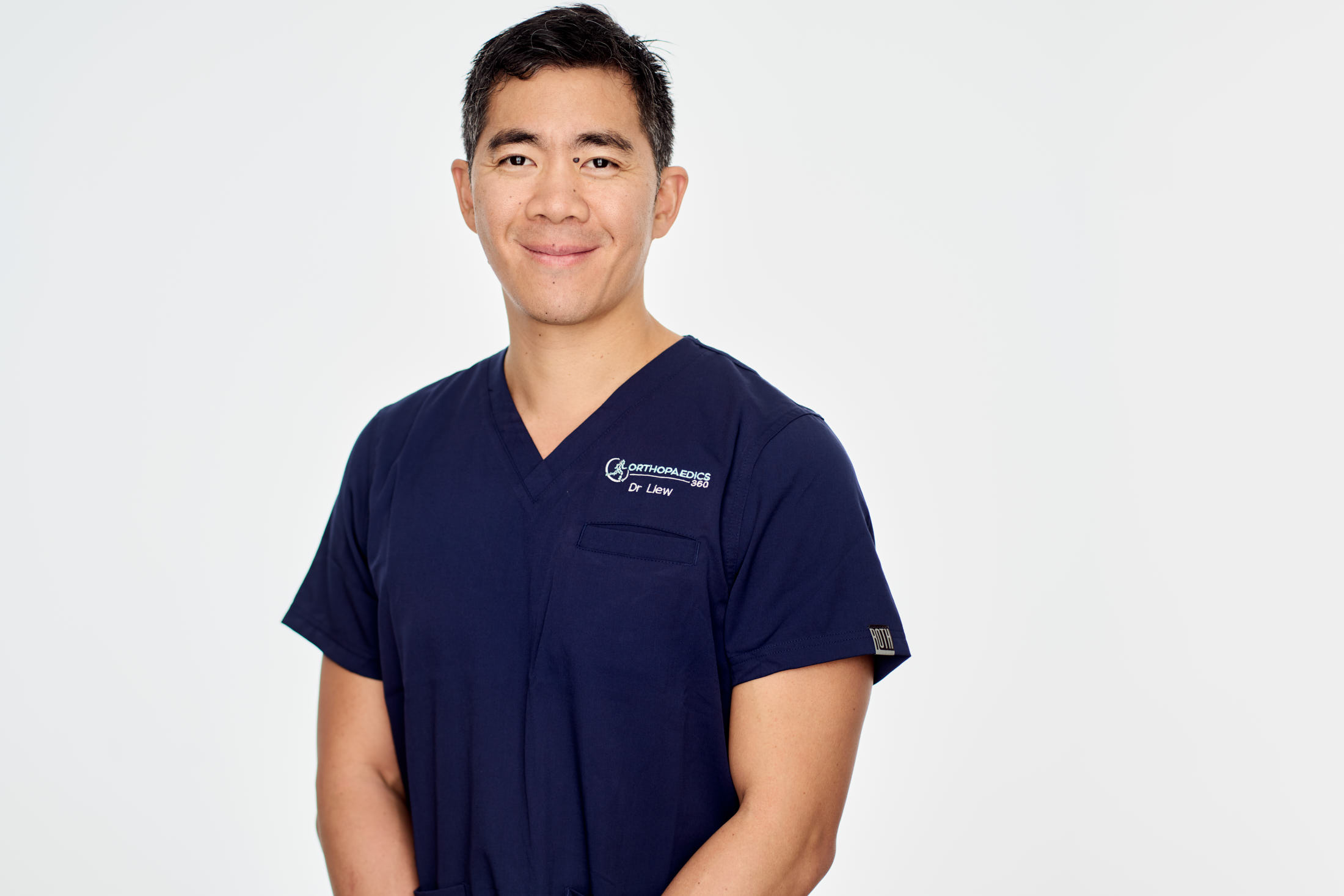
About Dr Chien-Wen Liew
Dedicated to Hip and Knee Replacements only


